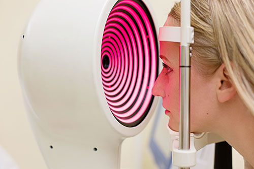
When patients come to an eye clinic to check their eyesight, they notice that doctors sometimes take colorful corneal photos called topography. What exactly is a corneal topography? Why are doctors interested in it?
First, let's talk about the cornea. The cornea is the most transparent layer of the eye. It has a certain curvature. The central thickness is about 0.5~0.55 mm, and the edge thickness is about 0.6~0.7 mm. Although it is very thin, it has a great effect and provides most of the refractive power for the eye. The shape of the cornea is different for each of us, and even the shape of cornea between two eyes of the same person is not the same. 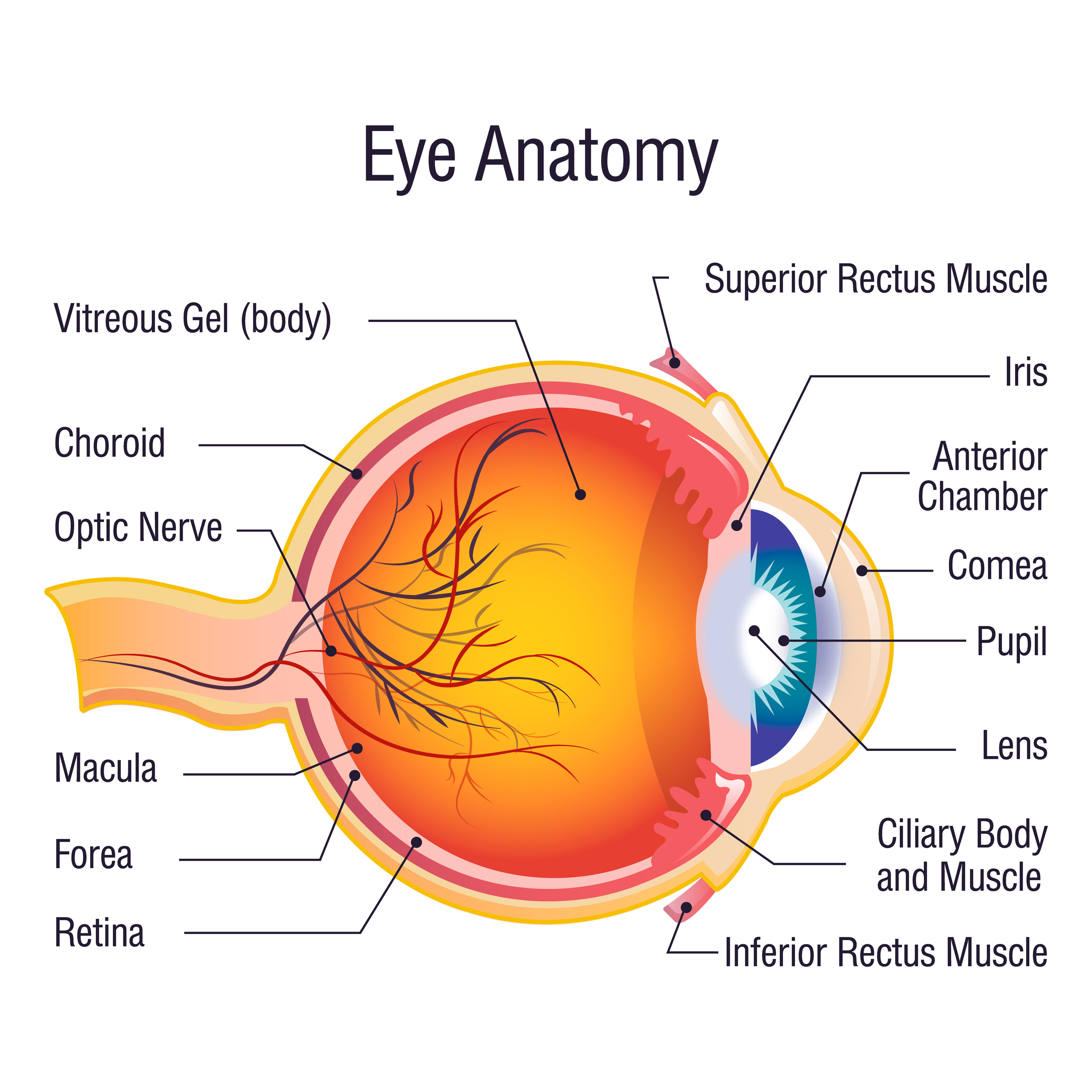 When the shape of the cornea does not match the overall structure of the eye, we will have refractive errors, which can be myopia, hyperopia or astigmatism. Therefore, it is very important to accurately determine the shape and curvature of the cornea. This can help to diagnose some eye diseases, such as keratoconus and the diagnostic accuracy is as high as 96% with this special tool.
The cornea is extremely thin and very sensitive. Before ophthalmic surgery or treatment, doctors need to pay extra attention to patient's corneal shape.
The corneal topography is an instrument that uses a computer-aided system to inspect the cornea. It can evenly project 28 or 34 rings on the cornea, from the centre of the cornea to the edge. With the help of a real-time image monitoring system, it photographs the cornea, and then uses a computer to analyse the graphics and data.
Just like the topographic maps on geography textbooks, there are also various colors on the corneal topography. Colors are used to represent areas that are "steep" or "flat" where red represents the steepest area and blue is for the flattest area. Therefore, the results displayed by the corneal topography are particularly intuitive and eye-catching.
Laser corneal specialists and doctors who fit RGP lenses, eg, ortho-k lenses are most interested in the shape of the cornea.
Most important clinical applications of corneal topography.
1: Detection of corneal pathologic conditions, most importantly keratoconus and pellucid marginal degeneration.
The detection of keratoconus is particularly important in patients who plan to undergo refractive surgery. Corneal topography can detect changes suggestive of early keratoconus in many patients without classic clinical manifestations of this disease, which are often classified as subclinical keratoconus.
2: Evaluation of irregular astigmatism, especially after penetrating keratoplasty.
Corneal topography is mostly valuable for detection of postoperative astigmatism, planning of removal of sutures, and postoperative fitting of RGP lenses.
3: Ortho-K lenses are special RGP lenses which are tailored to the shape of the cornea, therefore the requirements for corneal topography are higher.
The topographic map plays a decisive role in the success of the fitting. After wearing the OK lenses, the doctor will repeat the corneal topographic measurement to confirm the shaping effect.
It must be said that the corneal topography gives clinicians the insight into the corneal morphology. Needless to say that interpreting corneal topography is a matter of learning. Even a very senior doctor must be trained and practised for a long time to be proficient in the use of the equipment.
Copyright © Myopia Control Centre | Ortho-K Melbourne. All rights reserved